
Extracorporeal Shockwave Therapy for Persistent Achilles Tendinopathy: Why Getting the Diagnosis Right Comes First
Persistent Achilles pain is one of the most frustrating injuries to live with. It rarely responds overnight, it flares with activity you thought was safe, and by the time most people seek treatment they’ve already tried rest, stretching, and maybe an anti-inflammatory or two without lasting relief. Extracorporeal shockwave therapy (ESWT) has become one of the most researched non-surgical options for this problem – but it only works well when it’s used on the right condition. That’s the part we want to focus on in this post.
What is extracorporeal shockwave therapy?
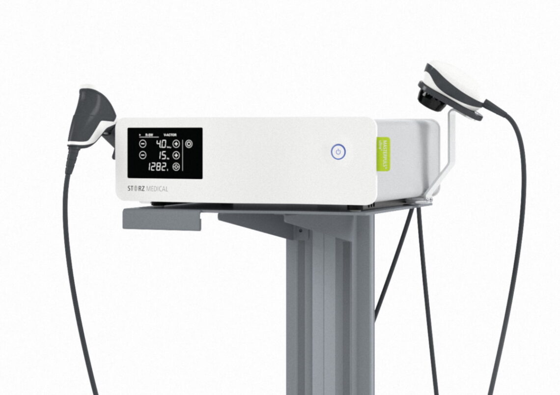
ESWT delivers acoustic pressure waves through the skin to the injured area of tendon. Two forms exist: focused shockwave, which concentrates energy at a specific depth, and radial shockwave, which disperses energy more broadly from the point of contact and is commonly used for superficial tendon and soft-tissue conditions like Achilles tendinopathy.
The working theory is that controlled mechanical stress to the tendon stimulates blood flow, disrupts abnormal pain signalling, and encourages a healing response in tissue that has become degenerative rather than simply inflamed – which is the case in most long-standing (“chronic”) tendinopathy.
Does it actually work for Achilles tendinopathy?
The evidence base is reasonably strong, though not uniformly one-sided.
A 2022 systematic review of randomised controlled trials looked at ESWT for both mid-portion and insertional Achilles tendinopathy and found it produced meaningful improvements in pain and function compared with standard care, particularly when added on top of a structured loading/exercise program.
Earlier placebo-controlled work has been more mixed. A double-blind trial of 43 patients with chronic Achilles tendinopathy found that both the real shockwave group and the sham shockwave group improved over the follow-up period, with pain scores dropping from roughly 7.5 to 3 in the treated group and from about 7.7 to 4.3 in the sham group – a reminder that placebo response and natural history both play a role in tendinopathy trials, and that ESWT tends to perform best as part of a broader rehabilitation plan rather than as a stand-alone fix.
More recent trial data, including a 2025 randomised controlled trial on insertional Achilles tendinopathy conducted in a private clinical setting, has specifically examined radial shockwave against sham treatment for pain, function, and quality-of-life outcomes over 12 weeks, reflecting how the research has matured toward answering exactly the question patients ask: is the real treatment better than doing nothing?
Taken together, the current picture is that ESWT has a reasonable evidence base for mid-portion and insertional Achilles tendinopathy, works best combined with eccentric loading exercise, and is not a guaranteed fix for everyone – which is exactly why case selection matters so much.
Why we scan before we treat
“Achilles pain” is not one diagnosis. Several distinct conditions live in the same small patch of anatomy at the back of the heel, and they can feel remarkably similar to the patient while requiring completely different management:
- Mid-portion or insertional Achilles tendinopathy – degenerative change within the tendon itself
- Retrocalcaneal bursitis – inflammation of the fluid-filled sacs near the tendon’s attachment
- Partial tendon tears – structural disruption of tendon fibres, sometimes with no dramatic injury moment
- Haglund’s deformity, enthesitis, or paratenonopathy – bone or sheath-related contributors to posterior heel pain
A narrative review of diagnostic ultrasound for Achilles-region conditions makes the point plainly: increasing use of imaging has helped confirm some of these diagnoses while also revealing that conditions such as retrocalcaneal bursitis and calcific tendinopathy get misclassified when clinicians rely on clinical examination alone. Identifying the correct cause of hindfoot pain matters because different conditions respond to entirely different treatments – some suited to medication, some to exercise, some to surgery.
This distinction isn’t academic when shockwave therapy is on the table. Applying mechanical shock energy to a bursa or to torn tendon fibres is a different proposition to applying it to degenerative but structurally intact tendon tissue. Research on partial tears has noted that their clinical presentation is often indistinguishable from tendinopathy or bursitis on examination alone, and that diagnostic ultrasound, colour Doppler, 0r MRI are needed to confirm what’s really going on. Case reports of tendon rupture following shockwave therapy in patients with pre-existing tendinopathy have also underlined that a prior diagnosis of tendinopathy is itself a risk factor for rupture – reinforcing why confirming tendon integrity beforehand, rather than assuming it, is the safer approach.
In short: the tools exist to tell these conditions apart, and using them changes what we recommend.
How this shapes our approach in clinic
Before we ever switch on a shockwave device, we perform a diagnostic scan to confirm exactly what we’re dealing with. In practice, that means:
- Confirming it’s genuinely tendinopathy – ruling out bursitis, paratenonopathy, or other enthesis-related contributors that need different treatment entirely.
- Checking tendon integrity – making sure there isn’t a partial tear that shockwave energy could aggravate rather than help.
- Locating the problem precisely – mid-portion and insertional tendinopathy don’t always respond to treatment in the same way, so knowing which one we’re treating, and exactly where, lets us target the shockwave application accurately.
- Setting expectations honestly – if imaging shows a tear or a condition shockwave isn’t suited to, we’ll tell you that and discuss what actually is appropriate, rather than defaulting to the treatment we happen to offer.
This is, admittedly, an extra step compared with clinics that go straight to treatment based on symptoms and a physical exam. We think it’s the right one. Shockwave therapy has real evidence behind it for the condition it’s designed to treat – but “the condition it’s designed to treat” is a specific, confirmable diagnosis, not just heel pain in general.
What to expect if you’re a candidate
If imaging confirms mid-portion or insertional Achilles tendinopathy without a tear, a typical course involves several sessions of radial shockwave delivered roughly weekly, usually paired with a structured eccentric or heavy-slow-resistance loading programme – the combination the evidence supports best. Mild soreness or redness after treatment is common and expected; a lasting increase in pain or new mechanical symptoms is not, and would prompt us to reassess.
The takeaway
Extracorporeal shockwave therapy has a solid, if not perfect, evidence base for chronic Achilles tendinopathy, and it works best as part of a broader rehab plan rather than in isolation. But “shockwave for Achilles pain” is only a sound recommendation once bursitis, tears, and other look-alike conditions have been ruled out. That’s why a diagnostic scan comes first in our clinic – not as an upsell, but because it’s what determines whether shockwave is actually the right tool for your tendon.
If you’ve had persistent Achilles pain that hasn’t settled with rest or basic exercises, get in touch – we’ll start with a proper diagnostic assessment so any treatment plan is based on what’s actually going on, not a guess.
MORE INFORMATION ABOUT SHOCKWAVE
References
- Paantjens MA, Helmhout PH, Backx FJG, van Etten-Jamaludin FS, Bakker EWP. Extracorporeal Shockwave Therapy for Mid-portion and Insertional Achilles Tendinopathy: A Systematic Review of Randomized Controlled Trials. Sports Medicine – Open. 2022;8:63. https://www.ncbi.nlm.nih.gov/pmc/articles/PMC9106789/
- Vahdatpour B, Forouzan H, Momeni F, Ahmadi M, Taheri P. Effectiveness of extracorporeal shockwave therapy for chronic Achilles tendinopathy: A randomized clinical trial. J Res Med Sci. 2018;23:37. https://pmc.ncbi.nlm.nih.gov/articles/PMC5961283/
- Alsulaimani B, Perraton L, Vallance P, Powers T, Malliaras P. Does shockwave therapy lead to better pain and function than sham over 12 weeks in people with insertional Achilles tendinopathy? A randomised controlled trial. Clin Rehabil. 2025;39(2):174–186. https://www.ncbi.nlm.nih.gov/pmc/articles/PMC11846266/
- Chimenti RL, et al. A Narrative Review of the Classification and Use of Diagnostic Ultrasound for Conditions of the Achilles Tendon. Diagnostics. 2020;10(11):944. https://www.ncbi.nlm.nih.gov/pmc/articles/PMC7696236/
- Minimum 3.5-year outcomes of operative treatment for Achilles tendon partial tears in the midportion and retrocalcaneal area. Arch Orthop Trauma Surg. https://www.ncbi.nlm.nih.gov/pmc/articles/PMC7488251/
- A case report of Achilles tendon distractive rupture after shock wave therapy. https://www.ncbi.nlm.nih.gov/pmc/articles/PMC11971564/
- MRI of the Achilles tendon- A comprehensive pictorial review. Part one. https://www.ncbi.nlm.nih.gov/pmc/articles/PMC8039565/
Note: this article is for general information and is not a substitute for individualised medical assessment.


